The mesothelium is a thin, protective layer of specialized cells that lines the internal organs and body cavities. The main functions of the mesothelium include:
- Protect internal organs from friction and injury.
- Produce a lubricating fluid that allows organs (like the lungs or intestines) to move smoothly against nearby structures.
- Support immune response, as mesothelial cells can react to inflammation and injury.
It is found in:
- Pleura: Covers the lungs and lines the chest cavity.
- Peritoneum: Lines the abdominal cavity and covers abdominal organs.
- Pericardium: Covers the heart and lines the surrounding cavity.
- Tunica vaginalis: Covers the testes.
Each of these areas has its own name and function, but all are lined by mesothelial cells. However, mesothelium is a thin lining and spreads over a large area. So, it is prone to malignant changes, which cause serious diseases, like mesothelioma.
What is Mesothelioma?
Mesothelioma is a rare and aggressive type of cancer that forms in the mesothelium. The most common type is pleural mesothelioma, which affects the lining around the lungs. Other less common forms include peritoneal mesothelioma, pericardial mesothelioma, and testicular mesothelioma.
The main cause of mesothelioma is exposure to asbestos, a naturally occurring mineral once widely used in construction, insulation, shipbuilding, automotive parts, and many industrial products.
When asbestos-containing materials are disturbed (during mining, construction, or demolition), they release tiny fibers into the air. These fibers can be inhaled or swallowed.
Once inside the body, the fibers can become lodged in the lungs, abdominal lining, or other tissues. The body cannot easily remove them.
As a result, over many years (usually 20–50), the fibers cause chronic irritation, scarring, and cellular damage. This can lead to DNA mutations and uncontrolled cell growth, which results in mesothelioma.
Mesothelioma is often difficult to diagnose in its early stages due to its non-specific symptoms and resemblance to other malignancies.
How is Mesothelioma Detected?
To diagnose mesothelioma, researchers rely on immunohistochemical markers – proteins that are specifically expressed in specific cell types.
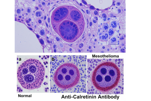
During mesothelioma, the highly expressed protein in mesothelial cells is calretinin. It is a calcium-binding protein found in neurons and mesothelial cells. Its primary function includes regulating calcium signaling and cell function.
Since calretinin is strongly expressed in mesothelial cells but is rarely found in other cancer cells, such as adenocarcinomas, it makes calretinin a highly specific biomarker for identifying mesothelial origin in cancer diagnosis.
So, researchers detect the amount of calretinin using the IHC technique. In this technique, researchers use an anti-calretinin antibody to identify mesothelioma.
What is Anti-Calretinin Antibody?
The anti-calretinin antibody is a lab-generated antibody that binds specifically to calretinin proteins in tissue samples.
Anti-Calretinin Antibodies:
- Have high diagnostic accuracy
- Are cost-effective and quick
- Works across different subtypes
It is used in immunohistochemistry (IHC) to detect the presence of calretinin in cells under a microscope.
How It Works:
- A tissue sample (typically from a biopsy) is placed on a slide.
- It is treated with the anti-calretinin antibody, which binds to calretinin if present.
- A staining reaction occurs, making the calretinin protein visible under a microscope.
- A positive stain (especially in both the nucleus and cytoplasm) confirms the presence of mesothelial cells, helping support a diagnosis of mesothelioma.
How Anti-Calretinin Antibody Helps in Mesothelioma Detection?
Differentiation from Other Malignancies
The anti-calretinin antibody plays a vital role in differentiating mesothelioma from metastatic adenocarcinoma. While both can appear in the pleural or peritoneal areas, their origins and treatment approaches are completely different.
Calretinin is rarely expressed in adenocarcinoma cells but is strongly expressed in mesothelial cells. So, a positive calretinin stain, especially when both the nucleus and cytoplasm are stained, means the presence of mesothelioma.
Part of an Immunohistochemical Panel
Although calretinin is highly sensitive and specific for mesothelial cells, it is not used alone. It is part of a panel of antibodies that pathologists use for accurate diagnosis. Commonly paired markers include:
- Positive mesothelial markers: Calretinin, WT-1, D2-40, CK5/6
- Negative mesothelial markers (positive in adenocarcinomas): CEA, TTF-1, Ber-EP4, MOC-31
This panel helps eliminate diagnostic uncertainty by showing a pattern of protein expression unique to mesothelioma.
Sensitivity and Specificity
Studies have shown that calretinin has a sensitivity of approximately 85–100% and a specificity of 75–95% for mesothelioma. This means it correctly identifies the disease in most patients while avoiding false positives in those without the disease.
Moreover, the nuclear and cytoplasmic staining pattern also adds to its diagnostic value compared to other markers that only show cytoplasmic staining.
The Bottom Line
The anti-calretinin antibody plays a central role in diagnosing mesothelioma. By specifically binding to calretinin, it helps pathologists differentiate mesothelioma from other malignancies, especially adenocarcinomas.
When used as part of an immunohistochemical panel, it provides high sensitivity and specificity, supporting timely and accurate diagnosis. So, it is a powerful tool when it comes to mesothelioma diagnosis.